count - a numerical computation or indication.

Addis count, a formerly common method of determining the number of red blood cells, white blood cells, epithelial cells, and casts, as well as the protein content in an aliquot of a twelve-hour urine specimen, used in the diagnosis and management of kidney disease.
Arneth count, a method of determining what percentages of neutrophils in a population have each number of nuclear lobes or segments between one and five; normally over 75 per cent should have two or three lobes. An increase in the percentages with fewer lobes, called a shift to the left, is seen in bacterial infections, whereas an increased number with hypersegmentation of the nuclei, called a shift to the right, is seen in cobalamin and folate deficiency. Called also neutrophil lobe c.
blood count, blood cell count, determination of the number of formed elements in a cubic millimeter of blood; it may be a complete blood count or it may measure just one of the formed elements. Methods include manual counts with a hemacytometer and automated counts with a flow cytometer, a Coulter counter, or other means.
complete blood count, a series of tests of the peripheral blood, including the erythrocyte count, erythrocyte indices, leukocyte counts, and sometimes platelet count.
differential blood count, differential leukocyte count, a leukocyte count that calculates the percentages of different types of cells. Cf. total leukocyte c.
erythrocyte count, determination of the number of erythrocytes in a unit volume of blood that has been diluted in an isotonic solution, done with an automatic counter such as a flow cytometer. Called also red blood cell c. and red cell c.
filament-nonfilament count, in the differential leukocyte count, determination of the number of juvenile and mature leukocytes.
leukocyte count, determination of the number of leukocytes in a unit volume of blood, usually after the erythrocytes have been lysed and the blood has been diluted; it may be done either manually with a hemacytometer or electronically. See total leukocyte c. and differential leukocyte c. Called also white blood cell c. and white cell c.
neutrophil lobe count, Arneth c.
platelet count, determination of the number of platelets per cubic millimeter of blood; it may be either a direct platelet count with a hemacytometer and a microscope or an indirect platelet count in which the ratio of platelets to erythrocytes on a blood smear is determined and the number of platelets is computed from the erythrocyte count.
red blood cell count, red cell count, erythrocyte c.
reticulocyte count, a calculation of the number of reticulocytes in 1 mm3 of peripheral blood, recorded as an absolute number or as the percentage of the erythrocyte count. It provides a means of assessing the erythropoietic activity of the bone marrow.
total leukocyte count, a leukocyte count measuring the total number of all the types in a given volume of blood. Cf. differential leukocyte c.
total lymphocyte count, a test that may be useful as an indicator of nutritional status and outcome.
white blood cell count, white cell count, leukocyte c.

Differential blood count -Whole #s or % - all=100
WBC count include:
polymorphonuclear neutrophils - PMNs, polys, segs (segmented neutrophils)
band neutrophils (bands, stabs)
lymphocytes (lymphocytes)
eosinophils (e-os)
basophils (bas-os)
monocytes (mono-s)
BLOOD GROUPS
1. an allotype (or phenotype) of erythrocytes defined by one or more cell surface antigens that are under the control of allelic genes. Antigenic determinants irregularly incite allotypic and sometimes xenotypic immune responses. Human blood groups are identified by agglutination supported by specific human or animal antisera and by lectins extracted from certain plants. An abbreviated classification of human blood groups is given in the accompanying table. 2. any of certain other characteristics or traits of a cellular or fluid component of blood, considered as the expression (phenotype or allotype) of the actions and interactions of dominant genes; used in medicolegal and other studies of human inheritance. Such characteristics include the antigenic groupings of erythrocytes, leukocytes, platelets, and plasma proteins. Called also blood type.
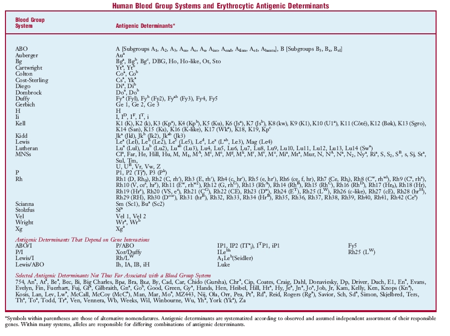
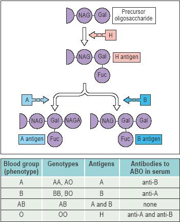
ABO blood group, the major human blood group system, dependent on the presence or absence of A and B antigens, which are largely glycolipids on the cell membrane. The gene for A is responsible for synthesis of N-acetyl-α-d-galactosaminyl transferase, whereas that for B is responsible for α-d-galactosyl transferase. Either A or B is created when one of these hexasaccharides is positioned by a specific transferase in 1→3 linkage to the β-d-galactose of an H-active oligosaccharide. Type O occurs when neither transferase is present or, very rarely (Bombay phenotype), when H antigen does not exist. When both transferases are present, type AB results. Differences in degree of transferase activity are determined at the same locus: weak transferase gives rise to weak antigens (A2, A3Ax, B3Bx). Similar oligosaccharides, especially in bacterial cell walls, immunize persons lacking A or B so that their serum contains anti-A or anti-B activity. A and B antigens are on the mucopolysaccharides of secretors; persons with dominant genes have H-active mucoids.
Auberger blood group, a blood group consisting of the erythrocytic antigen Aua, related to the Lutheran blood group.
Bg blood group, a blood group consisting of the erythrocytic human leukocyte antigens Bga, Bgb, Bgc, DBG, Ho, Ho-like, Ot, and Sto.
Cartwright blood group, Yt b. g.
Chido-Rodgers blood group, a blood group consisting of antigens Cha and Rga, antigenic determinants of fragments of the C4 component of complement.
Colton blood group, a blood group consisting of erythrocytic antigens Coa and Cob.
Cromer blood group, a blood group consisting of erythrocytic antigens Cra, Tca, Tcab, Dra, Esa, WESb, UMC, and IFC, which are located on the membrane protein called decay accelerating factor.
Diego blood group, a blood group consisting of the erythrocytic antigens Dia and Dib, determined by allelic genes. Dia is most frequent in South American Indians, Japanese, and Chinese.
Dombrock blood group, a blood group consisting of the erythrocytic antigens Doa and Dob, most common in people of European descent.
Duffy blood group, a blood group consisting principally of the erythrocytic antigens Fya and Fyb, determined by allelic genes. Amorphic genes are common in individuals of African descent.
Gerbich blood group, a blood group consisting of the erythrocytic antigens Ge 1, Ge 2, and Ge 3; although rare in most parts of the world, it has been found often in Papua New Guinea.
H blood group, a blood group consisting of antigen H; see also Bombay phenotype.
high frequency blood group, a group containing over 99 per cent of individuals, who have a type of erythrocyte antigens called public antigens.
Ii blood group, a high frequency blood group involving receptors of most cold reactive hemagglutinins; it is expressed most strongly on cord blood cells.
Kell blood group, a blood group consisting of multiple erythrocytic antigens, especially three pairs of alternates, determined by complex genes at one locus, including an amorph; also regulated by the X chromosome, it is associated with sex-linked chronic granulomatous disease. One antigen, K6, is more frequent in people of African descent.
Kidd blood group, a blood group consisting principally of Jka and Jkb antigens, determined by allelic genes; amorphic genes are most common in those of East Asian descent.
Knops blood group, a blood group consisting of antigens Kna, Knb, McCa, Sla, and Yka, which are located on complement receptor type 1.
Lan blood group, a blood group consisting of the erythrocytic antigen Lan.
Lewis blood group, a blood group determined by plasma glycolipids that adhere to erythrocytic surfaces. It is based on dominant independent Le genes, but interacts with the H precursor oligosaccharides of A and B. Whereas le/le provides the “double negative” blood type Le(a−b−), Le without H gives rise to Lea, i.e., blood type Le(a+b−), and that with H gives rise to LebH, i.e., blood type Le(a−b+).
low frequency blood group, any small group that has erythrocytic antigens found in fewer than 1 per cent of the population (private antigens).
Lutheran blood group, a complex blood group system consisting of antigens Lua and Lub; it somewhat resembles the Kell group in having pairs of alternative antigens and amorphic genes, but is also subject to a dominant independently segregating repressor.
MNSs blood group, a complex blood group system consisting principally of two pairs of antigens determined by closely linked genes (crossovers have been observed, but rarely). M and N, determined by allelic genes, depend on sialic (neuraminic) acid residues. S and s are also determined by allelic genes, and an amorphic gene is common in blacks when another antigen (U) is missing. The system also includes numerous low frequency antigens.
P blood group, a blood group system originally consisting of only P (now P1) antigen, but later found to include P2 (Tja), a very high frequency antigen, and P3 (PK), a very low frequency antigen. P1 is most common in people of African descent (90 per cent), less so in those of European descent (75 per cent), and least in those of East Asian descent (30 per cent).
Rh blood group, the most complex of all human blood groups because the genes differ by determining different numbers of antigens (Rh factors) and do so with remarkably different quality; over 40 antigens have been described to date. People of African descent show the greatest degree of diversity and East Asians the least. The major antigen, Rh1 (Rh0, D, or Rh0D), is highly immunogenic and before the development of passive immunization prophylaxis it was responsible for serious hemolytic disease of the newborn. Two other pairs of alternative antigens are inherited with or without Rh1; these are Rh21 (rhG or CG) and Rh4 (hr´ or c), and Rh3 (rh´ or E) and Rh5 (hr´ or e). The most common groups of antigens are R−1,−3,−21 (in Caucasians), R1,−3,−21 (in blacks), R1,−3,21 (in East Asians and Caucasians), and R1,3,−21 (in East Asians and Caucasians). Another antigen Rh10 (hrv, V) is common in blacks.
Scianna blood group, a blood group consisting of erythrocytic antigens Sc1 (formerly Sm) and Sc2 (formerly Bua).
Sid blood group, a blood group consisting of those with extra amounts of the public erythrocytic antigen Sda, referred to as Sd(a++).
Vel blood group, a blood group consisting of the erythrocytic antigens Vel 1 and Vel 2.
Wright blood group, a blood group consisting of the erythrocytic antigens Wra and Wrb.
Xg blood group, a blood group consisting of erythrocytic antigen Xga, which is determined by a gene on the long arm of the X chromosome.
Yt blood group, a blood group consisting of the erythrocytic antigens Yta and Ytb. Called also Cartwright b. g.
blood pressure, the pressure of blood against the walls of any blood vessel.the pressure of the blood on the walls of the arteries, dependent on the energy of the heart action, the elasticity of the walls of the arteries, and the volume and viscosity of the blood. The maximum or systolic blood pressure occurs near the end of the stroke output of the left ventricle of the heart. The minimum or diastolic blood pressure occurs late in ventricular diastole. Mean blood pressure is the average of the blood pressure levels, and basic blood pressure is that during quiet rest or basal conditions. See also hypertension and hypotension.
reported in mmHg or mm Hemoglobin, 
diastole
the dilatation, or period of dilatation, of the heart, especially of the ventricles; it coincides with the interval between the second and the first heart sounds. Cf. systole and see illustration at cardiac cycle, under cycle.
electrical diastole, that time during which the cell rests; it is represented by phase 4 of the action potential.
output
the yield; the total of anything produced by any functional system of the body.
basal acid output, on the pentagastrin test, the output of gastric acid for one hour with minimal stimulation in a fasting patient, expressed as mmol/hr.
cardiac output, the effective volume of blood expelled by either ventricle of the heart per unit of time (usually volume per minute); it is equal to the stroke volume multiplied by the heart rate.
energy output, the energy a body is able to manifest in work or activity.
maximal acid output, on the pentagastrin test, the output of gastric acid for one hour after administration of pentagastrin, expressed as mmol/hr.
peak acid output, on the pentagastrin test, after administration of pentagastrin, the sum of the two highest 15-minute outputs of gastric acid multiplied by 2, expressed as mmol/hr.
stroke output, see under volume.
urinary output, the amount of urine excreted by the kidneys.
work output of the heart, the amount of energy that the heart converts to work; usually expressed per heart beat (stroke work o.) or per minute (minute work o.).
hypotension
abnormally low blood pressure; seen in shock but not necessarily indicative of it.
chronic idiopathic orthostatic hypotension, chronic orthostatic hypotension, idiopathic orthostatic hypotension, Shy-Drager syndrome.Bradbury-Eggleston syndrome.
intradialytic hypotension, hypotension sometimes seen as a complication of hemodialysis.
orthostatic hypotension, postural hypotension, a fall in blood pressure associated with dizziness, blurred vision, and sometimes syncope, occurring upon standing or when standing motionless in a fixed position; it can be acquired or idiopathic, transient or chronic, and may occur alone or secondary to a disorder of the central nervous system such as the Shy-Drager syndrome.
supine hypotension, see under syndrome.
vascular hypotension, severe hypotension from dilatation of the blood vessels.
hypertension
high arterial blood pressure; various criteria for its threshold have been suggested, ranging from 140 mm Hg systolic and 90 mm Hg diastolic to as high as 200 mm Hg systolic and 110 mm Hg diastolic. Hypertension may have no known cause (essential or idiopathic h.) or be associated with other primary diseases (secondary h.).
accelerated hypertension, progressive hypertension marked by the funduscopic vascular changes of malignant hypertension but without papilledema.
adrenal hypertension, hypertension caused by an adrenal tumor that secretes mineralocorticoids, such as in hyperaldosteronism; in many cases it may be associated with excessive production of other adrenocortical hormones normally made in minute amounts.
benign intracranial hypertension, pseudotumor cerebri.
borderline hypertension, a condition in which the arterial blood pressure is sometimes within the normotensive range and sometimes within the hypertensive range; called also labile h.
chronic thromboembolic pulmonary hypertension, persistent pulmonary hypertension due to obstruction of a major pulmonary artery by an unresolved embolus.
essential hypertension, hypertension occurring without discoverable organic cause; called also primary h. and idiopathic h.
familial dyslipidemic hypertension, an inherited syndrome of seriously disordered blood lipid levels and essential hypertension.
gestational hypertension, pregnancy-induced h.
Goldblatt hypertension, hypertension experimentally induced with clamping that causes a Goldblatt kidney; called also Goldblatt phenomenon.
idiopathic hypertension, essential h.
idiopathic intracranial hypertension, pseudotumor cerebri.
intracranial hypertension, increased intracranial pressure; if symmetrically distributed it may have few neurologic symptoms (see pseudotumor cerebri), but if it is asymmetrical, as with hydrocephalus, neurologic symptoms are often severe.
labile hypertension, borderline h.
low-renin hypertension, essential hypertension associated with low levels of plasma renin concentration or low renin activity.
malignant hypertension, a severe hypertensive state with poor prognosis; it is characterized by papilledema of the ocular fundus with vascular exudative and hemorrhagic lesions, medial thickening of small arteries and arterioles, and left ventricular hypertrophy. Diastolic pressures as high as 130 mm Hg or more are commonly present.
ocular hypertension, persistently elevated intraocular pressure in the absence of any other signs of glaucoma; it may or may not progress to open-angle glaucoma.
persistent pulmonary hypertension of the newborn, a condition in newborns in which blood continues to flow through the foramen ovale and a patent ductus arteriosus, bypassing the lungs and resulting in hypoxemia. Called also persistent fetal circulation.
portal hypertension, abnormally increased blood pressure in the portal venous system, a frequent complication of cirrhosis.
hypertension of pregnancy, pregnancy-induced hypertension, the spectrum of preeclampsia and eclampsia.
primary hypertension, essential h.
pulmonary hypertension, pulmonary arterial hypertension, increased pressure (above 30 mm Hg systolic and 12 mm Hg diastolic) within the pulmonary arterial circulation.
renal hypertension, hypertension due to or associated with renal disease with a factor of parenchymal ischemia.
renovascular hypertension, hypertension due to occlusive disease of the renal arteries such as renal artery stenosis or fibromuscular dysplasia.
secondary hypertension, hypertension due to or associated with a variety of primary diseases, such as renal disorders, disorders of the central nervous system, endocrine diseases, and vascular diseases.
splenoportal hypertension, obstruction of the splenic venous system resulting in enlargement of the liver and manifestation of ascites and other evidence of portal cirrhosis.
symptomatic hypertension, hypertension accompanied by symptoms such as dizziness or headache.
systemic venous hypertension, elevation of systemic venous pressure, usually detected by inspection of the jugular veins.
vascular hypertension, hypertension.

FACTORS

factor
1. any of several substances or activities that are necessary to produce a result, e.g., a coagulation factor. Often, use of the term “factor” indicates that the chemical nature of the substance or its mechanism of action is unknown, as in endocrinology, where “factors” are renamed as “hormones” when their chemical nature is determined.

2. one of two or more quantities that multiplied together form a product.

3. a coefficient or conversion factor, a number by which a quantity is multiplied to produce a change of units of measurement.

factor I, see under
coagulation f's.
factor II, see under coagulation f's.
factor III, see under coagulation f's.
factor IV, see under coagulation f's.
factor V, see under coagulation f's.
factor VI, see under coagulation f's.
factor VII, see under coagulation f's.
factor VIII, see under coagulation f's.
factor IX, see under coagulation f's.
factor X, see under coagulation f's.
factor XI, see under coagulation f's.
factor XII, see under coagulation f's.
factor XIII, see under coagulation f's.
factor A, former name for the alternative pathway complement factor C3; see under complement.
accelerator factor, f. V; see under coagulation f's.
activation factor, f. XII; see under coagulation f's.
angiogenesis factor, a substance that causes the growth of new blood vessels, found in tissues with high metabolic requirements such as cancers and the retina; it is also released by hypoxic macrophages at the edges or outer surface of a wound and initiates revascularization in wound healing.
antigen-specific T-cell helper factor, a soluble factor produced by helper T cells that activates other lymphocytes that are specific for the stimulating antigen; it may itself bind antigen.
antigen-specific T-cell suppressor factor, a soluble factor produced by suppressor T cells following immunization; it produces antigen-specific suppression of the immune response and may itself bind antigen.
antihemophilic factor, f. VIII, see under coagulation f's.a preparation of factor VIII used in the treatment of hemophilia A, including preparations derived from human plasma (antihemophilic factor and cryoprecipitated antihemophilic factor) or porcine plasma (antihemophilic factor [porcine]) and those produced by recombinant technology (antihemophilic factor [recombinant]).a sterile freeze-dried powder containing the factor VIII fraction prepared from human venous plasma; used to arrest hemorrhage or to prevent hemorrhage during surgery or other procedures in patients with hemophilia A and in the treatment of von Willebrand disease; administered intravenously.
antihemophilic factor A, f. VIII; see under coagulation f's.
antihemophilic factor B, f. IX; see under coagulation f's.
antihemophilic factor C, f. XI; see under coagulation f's.
antihemophilic factor (human), antihemophilic f. (def. 3).
antihemophilic factor (porcine), a highly purified sterile freeze-dried concentrate of porcine coagulation factor VIII obtained from porcine plasma; it is less antigenic than human factor VIII and is used in the treatment of hemophilia A in patients with circulatory antibodies to factor VIII and in patients with spontaneously acquired inhibitors to human factor VIII; administered by intravenous infusion.
antihemophilic factor (recombinant), a sterile, purified, concentrate produced by recombinant technology using cultured hamster cell lines that have been transfected with a gene for human coagulation factor VIII, and having biological activity comparable to that of plasma-derived coagulation factor VIII; used to prevent or arrest hemorrhage during surgery or other procedures in patients with hemophilia A; administered intravenously.
antinuclear factor, see under antibody.
apoptotic protease activating factor 1, a cytoplasmic protein that is activated by cytochrome c (released from mitochondria) and deoxyadenosine triphosphate (dATP); it in turn binds to and activates caspase 9, initiating the apoptotic cascade.
atrial natriuretic factor, atrial natriuretic peptide.sometimes more specifically, the prohormone form of atrial natriuretic peptide.
autocrine growth factor, a polypeptide, produced endogenously by a cell free from usual external growth controls, that may act on its producer cell to cause malignant transformation.
autocrine motility factor, name given to glucose-6 isomerase in its identity as a stimulator of motility in cancer cells.
factor B, a complement component that participates in the alternative pathway of complement activation (see complement), binding to C3b to form C3bB, which is a substrate for factor D.
basophil chemotactic factor, a lymphokine produced by activated lymphocytes that is chemotactic for basophils, possibly responsible for the influx of basophils into sites of inflammation (Jones-Mote reaction).
B cell differentiation factors, factors derived from T cells that stimulate B cells to differentiate into antibody-secreting cells. Cf. B lymphocyte stimulatory f's.
B cell growth factors, factors derived from T cells that stimulate B cells to proliferate in vitro but (unlike B cell differentiation factors) do not stimulate antibody secretion. Cf. B lymphocyte stimulatory f's.
blastogenic factor, lymphocyte mitogenic f.
B lymphocyte stimulatory factors, a system of nomenclature for factors that stimulate B cells, replacing individual factor names (e.g., B cell differentiation factor). Each factor is designated by BSF and a number, BSF1, BSF2, etc., with the letter p prefixed to the number for factors that have not been purified or whose structure has not been identified. BSF1 is interleukin-4 and BSF2 is interleukin-6.
bone factor, in periodontal disease, the systemic influence on alveolar bone loss in response to local inflammatory processes.
CAMP factor, see under test.
Castle factor, Castle intrinsic factor, intrinsic f.
chemotactic factor, a substance that induces chemotaxis. See chemoattractant and chemorepellent. Called also chemotaxin.
Christmas factor, f. IX; see under coagulation f's.
citrovorum factor, folinic acid.
clotting factors, coagulation factors.
C3 nephritic factor, an autoantibody that binds the alternative complement pathway C3 convertase C3b,Bb and prevents its inactivation by factor H, resulting in chronic fluid phase alternative pathway activation and complete consumption of plasma C3; found in the serum of many patients with type II membranoproliferative glomerulonephritis.
coagulation factors, substances in the blood that are essential to the clotting process and hence, to the maintenance of normal hemostasis. They are designated by Roman numerals, to which the notation “a” is added to indicate the activated state. Platelet factors (q.v.) also play a role in coagulation.
coagulation factor IX (human), a purified, sterile, dried concentrate of factor IX derived from pooled human plasma; used in the prophylaxis and treatment of bleeding in patients with hemophilia B, administered intravenously. Cf. coagulation f. IX (recombinant) and factor IX complex.
coagulation factor IX (recombinant), a sterile, dried concentrate of factor IX prepared by recombinant means; used in the prophylaxis and treatment of bleeding in patients with hemophilia B, administered intravenously. Cf. coagulation f. IX (human) and factor IX complex.
colony-stimulating factors, a group of glycoprotein lymphokine growth factors produced by blood monocytes, tissue macrophages, and stimulated lymphocytes; they are required for differentiation of stem cells into granulocyte and monocyte cell colonies, and were originally named according to their ability to stimulate the production of morphologically distinguishable myeloid cell colonies in vitro. In vivo, they stimulate the production of granulocytes and macrophages and enhance the actions of mature cells. Colony-stimulating factors can be produced by recombinant DNA techniques and have been used experimentally as anticancer agents and for the restoration of hematopoietic function after myelosuppression in transplantation and cancer chemotherapy.
contact factor, f. XII; see under coagulation f's.
cord factor, a mycoside produced by those strains of Mycobacterium tuberculosis that characteristically grow in long serpentine cords.
cryoprecipitated antihemophilic factor, a sterile, frozen concentrate of antihemophilic factor prepared from the coagulation factor VIII–rich cryoprotein fraction of human venous plasma; used for autologous replacement of factor VIII in patients with hemophilia A and for the treatment of von Willebrand disease, hypofibrinogenemia, and coagulation factor XIII deficiency, administered intravenously.
crystal-induced chemotactic factor, a glycoprotein produced by neutrophils upon ingestion of monosodium urate or calcium pyrophosphate crystals, which is directly chemotactic for neutrophils and is thought to be involved in the inflammatory process in gouty arthritis.
factor D, a serine protease of the alternative complement pathway that cleaves factor B bound to C3b. The reaction releases Ba while leaving Bb bound to C3b to form the C3bBb that is the C3 convertase of the alternative pathway.
decay accelerating factor, a protein of most blood cells as well as endothelial and epithelial cells, CD55; it protects the cell membranes from attack by autologous complement, either by preventing assembly of convertase on the cell surface or by accelerating its decay once bound.
diabetogenic factor, see under hormone.
diffusion factor, Duran-Reynals factor, hyaluronidase.
elongation factor, one of two soluble proteins (EF-1 and EF-2) involved in the addition of each amino acid to the growing polypeptide chain in protein synthesis (see translation).
endothelial-derived relaxing factor, endothelium-derived relaxing factor, nitric oxide.
eosinophil chemotactic factor, eosinophil chemotactic f. of anaphylaxis.a lymphokine produced by activated lymphocytes that is chemotactic for eosinophils.
eosinophil chemotactic factor of anaphylaxis, eosinophil chemoattractants released by basophils and mast cells in immediate hypersensitivity reactions. ECF-A activity is associated with two acidic tetrapeptides (Ala-Gly-Ser-Glu and Val-Gly-Ser-Glu) and with less well characterized larger peptides, which are chemotactic for eosinophils and, to a lesser degree, for neutrophils. Some ECF-A activity is due to arachidonic acid metabolites (leukotriene B, 12-HETE, and 12-HHT). Called also eosinophil chemotactic f. (ECF).
epidermal growth factor, a mitogenic polypeptide originally extracted from the submandibular glands of male mice; it is produced by many cell types and is made in large amounts by some tumors. It promotes growth and differentiation, is essential in embryogenesis, and is also important in wound healing. It has been found to be part of a family of compounds that includes human epidermal growth factor, transforming growth factors, and amphiregulin.
epidermal growth factor, human, a mitogenic polypeptide found in humans that is 70 per cent homologous with the epidermal growth factor of mice; it promotes growth and differentiation, is essential in embryogenesis, and is important in wound healing. It is produced by many normal cell types and is made in large amounts by some tumors; the kidneys are the major source of the circulating factor. Called also urogastrone.
epithelial growth factor, human epidermal growth f.
erythropoietic stimulating factor, erythropoietin.
extrinsic factor, vitamin B12 (cyanocobalamin).
F factor, fertility factor, F plasmid.
fibrin-stabilizing factor, f. XIII; see under coagulation f's.
fibroblast growth factor, see under hormone.
Fitzgerald factor, high-molecular-weight kininogen.
Fletcher factor, prekallikrein.
glass factor, f. XII; see under coagulation f's.
glucose tolerance factor, a biologically active complex of chromium and nicotinic acid that facilitates the reaction of insulin with receptor sites on tissues.
granulocyte colony-stimulating factor, a colony-stimulating factor, secreted by stimulated endothelial cells, fibroblasts, and macrophages, that stimulates the production of neutrophils from precursor cells. A recombinant form used therapeutically is called filgrastim.
granulocyte-macrophage colony-stimulating factor, a colony-stimulating factor, secreted by activated T lymphocytes and macrophages and by stimulated endothelial cells, that binds to a specific receptor found on stem cells and most myelocytes and stimulates their differentiation into granulocytes and macrophages. GM-CSF also enhances the function of mature granulocytes and macrophages in vitro. A recombinant form used therapeutically is called sargramostim.
growth factor, an imprecise term denoting any of numerous substances that promote normal or pathological growth of cells or tissue, including the epidermal growth factors, insulin-like growth factors, nerve growth factors, platelet-derived growth factors, and transforming growth factors.
factor H, a glycoprotein that binds to C3b (see under complement ) and acts as an alternative pathway complement inhibitor by interfering with the binding of factor B to C3b; it also acts as a cofactor in the conversion of C3b to the inactive form iC3b by factor I.
Hageman factor, f. XII; see under coagulation f's.
hematopoietic growth factors, a group of substances with the ability to support hematopoietic colony formation in vitro, including erythropoietin, interleukin-3, and colony-stimulating factors. All except erythropoietin stimulate mature cells, have overlapping capabilities to affect progenitor cells of several blood cell lines, and also affect cells outside the hematopoietic system.
hepatocyte growth factor, a potent mitogen and inducer of hepatocyte proliferation, produced by nonparenchymal cells in the liver and by mesenchymal cells in many other organs.
hepatocyte nuclear factor, any of a group of transcription factors of liver-specific genes involved in a variety of functions, including carcinogenesis, atherosclerosis, hyperlipidemia, insulin resistance, and hypertension.
high-molecular-weight neutrophil chemotactic factor, neutrophil chemotactic f.
histamine-releasing factor, a lymphokine, believed to be produced by macrophages and B lymphocytes, that induces the release of histamine by IgE-bound basophils occurring in late phase allergic reaction in sensitive individuals.
homologous restriction factor, a regulatory protein that binds to the membrane attack complex factor C8 of autologous cells and by preventing C9 insertion into the membrane and subsequent polymerization so inhibits the final stages of complement activation.
hydrazine-sensitive factor, former name for alternative pathway complement factor C3; see under complement.
hyperglycemic-glycogenolytic factor, former name for glucagon.the hyperglycemic component of growth hormone extracts.
factor I, a plasma enzyme that regulates both classical and alternative pathways of complement activation (see complement). In the classical pathway, it blocks the formation of C3 convertase by catabolizing C4b; in the alternative pathway, it inactivates C3b by cleaving it to form iC3b, requiring also a cofactor such as factor H, MCP (membrane cofactor protein), or CR1 (complement receptor 1).
immunoglobulin-binding factor, a lymphokine having the ability to bind IgG complexed with antigen and prevent complement activation, possibly Fc receptors shed from T cells.
inhibiting factors, factors elaborated by one structure (such as the hypothalamus) that inhibit release of hormones by other structures (such as the adenohypophysis). The term is applied to substances of unknown chemical structure; those of established chemical identity are called inhibiting hormones.
initiation factor, one of three soluble proteins (IF-1, IF-2, and IF-3) involved in the binding of mRNA and the first aminoacyl-tRNA to the small ribosomal subunit and the attachment of the small subunit to the large subunit at the beginning of protein synthesis (see translation).
insulinlike growth factors, serum peptides with insulin-like actions, formerly called somatomedins (q.v.). IGF-I (formerly somatomedin C) is an important growth hormone–dependent mediator of cell growth and replication. IGF-II (formerly somatomedin A) appears to be essential for normal embryonic development and may play special roles in the central nervous system. Both are similar in sequence and structure to proinsulin.
intestinal trefoil factor, trefoil f. 3.
intrinsic factor, a glycoprotein secreted by the parietal cells of the gastric glands, necessary for the absorption of vitamin B12 (cyanocobalamin). Lack of intrinsic factor, with consequent deficiency of vitamin B12, results in pernicious anemia.
labile factor, f. V; see under coagulation f's.
Laki-Lorand factor, f. XIII; see under coagulation f's.
LE factor, an antinuclear antibody present in the blood serum in systemic lupus erythematosus, having a sedimentation rate of 7S and reacting with leukocyte nuclei in the LE cell test.
leukocyte inhibitory factor, a lymphokine that inhibits the migration of polymorphonuclear leukocytes but not macrophages.
lymph node permeability factor, a vasoactive factor, distinct from histamine, serotonin, bradykinin, and kallikrein, that is released without immunologic stimulus from many tissues, including lymph nodes, spleen, kidney, liver, and muscle.
lymphocyte-activating factor, interleukin-1.
lymphocyte blastogenic factor, lymphocyte mitogenic f.
lymphocyte mitogenic factor, lymphocyte-transforming factor, a nondialyzable heat-stable macromolecule, mol. wt. approximately 20,000–30,000, released by lymphocytes stimulated by specific antigen, that causes nonstimulated lymphocytes to undergo blast transformation and cell division.
macrophage-activating factor, interferon-γ.
macrophage chemotactic factor, a lymphokine produced by activated lymphocytes that is chemotactic for macrophages.
macrophage colony-stimulating factor, a colony-stimulating factor secreted by macrophages, stimulated endothelial cells, and most tissues, that stimulates the production of macrophages from precursor cells and maintains the viability of mature macrophages in vitro.
macrophage-derived growth factor, a substance released by macrophages below the surface of a wound that induces the proliferation of fibroblasts, with consequent deposition of collagen, fibronectin, and glycosaminoglycan.
macrophage growth factor, any of various glycoproteins that permit macrophages harvested from peritoneal exudates to proliferate in liquid-suspension cultures and form colonies consisting solely of mononuclear phagocytes.
macrophage inhibition factor, macrophage inhibitory factor, migration inhibitory f.
migration inhibition factor, migration inhibitory factor, a lymphokine that inhibits macrophage migration.
mitogenic factor, lymphocyte mitogenic f.
müllerian duct inhibitory factor, müllerian inhibiting factor, müllerian regression factor, antimüllerian hormone.
myocardial depressant factor, a peptide putatively formed in response to a fall in systemic blood pressure related to sepsis; it has a negatively inotropic effect on myocardial muscle fibers.
necrotizing factor, necrotoxin.
nerve growth factor, a protein consisting of two identical polypeptide chains associated with two gamma subunits (enzymes) and two alpha subunits; first isolated from mouse sarcoma and later from snake venom and mouse salivary glands, it stimulates the growth of sensory and sympathetic nerve cells and of the adrenal medulla and has been found to be secreted by a variety of normal and neoplastic cells, including those in humans.
neutrophil chemotactic factor, a poorly characterized chemotactic factor, mol. wt. approximately 750,000, that attracts neutrophils but not eosinophils or monocytes and is released by basophils or mast cells in immediate hypersensitivity reactions. Called also high-molecular-weight neutrophil chemotactic f. (HMW-NCF).a lymphokine produced by activated lymphocytes that is chemotactic for neutrophils.
osteoclast-activating factor, a lymphokine that stimulates bone resorption; it is a small protein unrelated to parathyroid hormone and may be involved in the bone resorption associated with multiple myeloma and other hematologic neoplasms or inflammatory disorders such as rheumatoid arthritis and periodontal disease.
factor P, properdin.
pellagra-preventive factor, niacin.
platelet factors, factors important in hemostasis, contained in or attached to the platelets; they act together with coagulation factors.
platelet-activating factor, a substance released by basophils and mast cells in immediate hypersensitivity reactions and macrophages and neutrophils in other inflammatory responses; it is an extremely potent mediator of bronchoconstriction and of the platelet aggregation and release reactions. It differs from other known biochemical mediators in being a phospholipid. Called also PAF-acether or AGEPC (acetyl glyceryl ether phosphoryl choline).
platelet-derived growth factor, a substance found in the alpha granules of platelets, capable of inducing proliferation of vascular endothelial cells, vascular smooth muscle cells, fibroblasts, and glia cells; its action contributes to the repair of damaged vascular walls.
P.-P. factor, niacin.
prolactin-inhibiting factor, see under hormone.
prolactin-releasing factor, see under hormone.
Prower factor, f. X; see under coagulation f's.
R factor, R plasmid.
recruitment factor, lymphocyte mitogenic f.
releasing factors, factors elaborated in one structure (such as the hypothalamus) that effect the release of hormones from another structure (such as the adenohypophysis). The term is applied to substances of unknown chemical structure; those of established chemical identity are called releasing hormones (see under hormone).two soluble proteins (RF-1 and RF-2) involved in the release of the completed polypeptide chain from the ribosome when a termination codon is encountered during protein synthesis (see translation). RF-1 recognizes the termination codon UAA or UAG and RF-2 recognizes UAA or UGA.
resistance-inducing factor, see Rubin test (def. 2), under test.
resistance transfer factor, the portion of an R plasmid in a bacterial cell that contains the genes for conjugation and replication.
Rh factor, Rhesus factor, any of numerous antigens (agglutinogens) that may be present on the membrane of erythrocytes and that determine the Rh blood group system; the most common ones are called (in one system) Rh 1, Rh 3, Rh 4, Rh 5, and Rh 21. See Rh blood group, under blood group. Called also Rh antigen.
rheumatoid factor, antibodies directed against antigenic determinants, i.e., Gm, in the Fc region of IgG, found in the serum of about 80 per cent of patients with classical or definite rheumatoid arthritis but in only about 20 per cent of patients with juvenile rheumatoid arthritis; rheumatoid factors may be IgM, IgG, or IgA antibodies, although serologic tests measure only IgM. Rheumatoid factors also occur in other connective tissue diseases and infectious diseases (Sjögren syndrome, systemic lupus erythematosus, sarcoidosis, subacute bacterial endocarditis, infectious hepatitis, leprosy).
risk factor, a clearly defined occurrence or characteristic that has been associated with the increased rate of a subsequently occurring disease; causality may or may not be implied.
sex factor, F plasmid.
Simon septic factor, decrease of eosinophils and increase of neutrophils in the blood in pyogenic infections.
skeletal growth factor, a protein that stimulates growth of osteocytes.
skin reactive factor, a lymphokine derived from antigen-stimulated lymphocytes that augments delayed hypersensitivity skin reaction, increasing capillary permeability and infiltration of monocytes; perhaps a mixture of other lymphokines.
somatotropin release–inhibiting factor, somatostatin.
spreading factor, hyaluronidase.
stable factor, f. VII; see under coagulation f's.
steroidogenic factor 1, a zinc fingerorphan nuclear receptor expressed in primary steroidogenic tissues and important for the development of the adrenal glands and for the regulation of several genes involved in reproduction, steroidogenesis, and male sexual differentiation; SF-1 disruption results in adrenal and gonadal agenesis, absence of pituitary gonadotrophs, ambiguous genitalia, and abnormalities of the hypothalamus.
Stuart factor, Stuart-Prower factor, f. X; see under coagulation f's.
sulfation factors, former name for insulin-like growth f's.
sun protection factor, sun protective factor, the ratio between the number of minimal erythema doses required to induce erythema through a film of sunscreen and that for unprotected skin.
T-cell growth factor, former name for interleukin-2.
tissue factor, f. III; see under coagulation f's.
transcription factor, a protein that regulates gene expression by binding to a DNA sequence adjacent to a sequence encoding a protein or RNA product, thus switching the gene on or off.
transfer factor, a dialyzable extract obtained from lysates of peripheral blood lymphocytes that is capable of transferring antigen-specific cell-mediated immunity (delayed-type hypersensitivity) from donor to recipient and also has nonspecific immunostimulatory activity; it appears to contain both protein and RNA but not DNA and to consist of small molecules (mol. wt. less than 10,000). TF is nonantigenic and does not transfer humoral immunity. It has been used in the treatment of a variety of immunodeficiency diseases.elongation f.
transforming growth factor, either of two types of proteins secreted by transformed cells and stimulating growth of normal cells, although not causing transformation. TGF-α binds the epidermal growth factor receptor and also stimulates growth of microvascular endothelial cells. TGF-β comprises several proteins, which are found in hematopoietic tissue, stimulate wound healing, and in vitro are antagonists of lymphopoiesis and myelopoiesis. Similar proteins involved in inducing the change of undifferentiated tissues into specific types of tissues, including bone morphogenetic proteins, activins, and inhibins, are said to belong to the superfamily of TGF-β proteins.
trefoil factor, any of a family of protease-resistant peptides having a three-loop structure consisting of six cysteine residues and three disulfide bonds, expressed throughout the gastrointestinal tract. They promote epithelial cell migration and gastrointestinal wound healing, prevent apoptosis and anoikis, and contribute to mucosal protection; TFF1 is also a tumor suppressor. TFF1 is called also pS2; TFF2, spasmolytic polypeptide or spasmolysin; and TFF3, intestinal trefoil f.
tumor-angiogenesis factor, a factor produced by cancer cells of solid tumors that stimulates the growth of blood vessels into the tumor.
tumor necrosis factor, either of two lymphokines that are capable of causing in vivo hemorrhagic necrosis of certain tumor cells, but not affecting normal cells; they have been used as experimental anticancer agents but can also induce shock when bacterial endotoxins cause their release. Tumor necrosis factor α, formerly called cachectin, contains 157 amino acids and is produced by macrophages, eosinophils, and NK cells. Tumor necrosis factor β is lymphotoxin and contains 171 amino acids.
V factor, an accessory substance required for the growth of certain species of Haemophilus, replaceable by nicotinamide-adenine dinucleotide (NAD) or nicotinamide-adenine dinucleotide phosphate (NADP) and present in red blood cells. Cf. X f.
vascular endothelial growth factor, vascular permeability factor, a peptide factor, existing in four forms with different lengths (121, 165, 189, and 206 amino acids), that is mitogenic for vascularendothelial cells and promotes tissue vascularization; levels are elevated in hypoxia and it is important in tumor angiogenesis.
von Willebrand factor, a glycoprotein synthesized in endothelial cells and megakaryocytes that circulates complexed to factor VIII (see under coagulation f's); it mediates adhesion of platelets to damaged epithelial surfaces and may participate in platelet aggregation. Deficiency results in the prolonged bleeding time seen in von Willebrand disease. note: this factor was originally considered to be part of factor VIII, so that in older terminology the term factor VIII generally refers to the complex of the two factors.
X factor, an accessory substance required for the aerobic growth of certain species of Haemophilus replaceable by hemin or other iron porphyrin compounds, and present in red blood cells. It is heat stable and is not destroyed by autoclaving. Cf. V f.

TYPES

type -something with particular characteristics, such as a person, substance, or case of a disease. Cf. constitution and diathesis.

ampullary type of renal pelvis, a type of renal pelvis in which the calices open into a saclike central chamber.
asthenic type, a constitutional type marked by a slender body, long neck, long, flat chest and abdomen, and poor muscular development.
athletic type, a constitutional type marked by broad shoulders, deep chest, flat abdomen, thick neck, and good muscular development.
blood type, blood group; see under B.
body type, constitutional t.
branching type of renal pelvis, a type of renal pelvis without a central chamber, but simply formed by the merger of several calices at the opening into the ureter.
constitutional type, a constellation of traits related to body build.
dysplastic type, any constitutional type that differs from the asthenic, the athletic, and the pyknic types.
Fitzpatrick skin type, one of the six skin types according to the Fitzpatrick scale.
mating type, in ciliate protozoa, certain bacteria, and certain fungi, the equivalent of a sex; as many as eight sexes are present in some species of protozoa.
personality type, any of various categories of both normal and abnormal personality variants; usually they derive from a theory-based topology, such as introvert/extrovert or oral/anal/phallic.
phage type, an intraspecies type of bacterium demonstrated by phage typing (see under typing); called also phagotype.
pyknic type, a constitutional type marked by a rounded body, large chest, thick shoulders, broad head, and short neck.
sympatheticotonic type, a type of physical constitution characterized by sympathicotonia.
test type, see test type.
wild type, the typical form occurring in a natural population or in the standard laboratory stock, as a strain, phenotype, or gene, and therefore designated as representative of the group.


RECEPTORS

re·cep·tor

1. a molecular structure within a cell or on the cell surface and characterized by (1) selective binding of a specific substance and (2) a specific physiologic effect that accompanies the binding, e.g., membrane receptors for peptide hormones, neurotransmitters, antigens, complement fragments, and immunoglobulins, and nuclear receptors for steroid hormones.

2. a sensory nerve terminal that responds to stimuli of various kinds; classified in various ways including by the type of stimulus (see chemoreceptor, mechanoreceptor, photoreceptor, and thermoreceptor) and by the location in the body (see exteroceptor, interoceptor, and proprioceptor).


α-adrenergic receptors, adrenergic receptors that respond to norepinephrine and to such blocking agents as phenoxybenzamine and phentolamine. They are subdivided into two types: α1, found in smooth muscle, heart, and liver, with effects including vasoconstriction, intestinal relaxation, uterine contraction and pupillary dilation, and α2, found in platelets, vascular smooth muscle, nerve termini, and pancreatic islets, with effects including platelet aggregation, vasoconstriction, and inhibition of norepinephrine release and of insulin secretion. Called also alpha or alpha-adrenergic receptors.
adrenergic receptor, a site on an effector organ innervated by postganglionic adrenergic fibers of the sympathetic nervous system, classified as either α-adrenergic or β-adrenergic according to its reaction to norepinephrine and epinephrine, as well as to certain blocking and stimulating agents. Called also adrenoceptor and adrenoreceptor.
alpha receptors, alpha-adrenergic receptors, α-adrenergic r's.
β-adrenergic receptors, adrenergic receptors that respond particularly to epinephrine and to such blocking agents as propranolol. They are subdivided into two basic types: β1, found in the myocardium and causing lipolysis and cardiac stimulation, and β2, found in smooth and skeletal muscle and liver and causing bronchodilation, vasodilation, and increased presynaptic release of norepinephrine. A third type, β3, is atypical; it is more sensitive to norepinephrine than to epinephrine, is relatively resistant to propranolol blockade, and may be involved in lipolysis regulation in adipose tissue. Called also beta or beta-adrenergic r's.
B cell antigen receptors, monomeric IgM, IgD, and (on memory cells only) IgG that is attached to the cell membrane of B lymphocytes and which, in conjunction with T cell help, triggers B cell activation on contact with antigen.
beta receptors, beta-adrenergic receptors, β-adrenergic r's.
cell-surface receptor, membrane r.
cholinergic receptor, a type of cell-surface receptor that binds the neurotransmitter acetylcholine and mediates its action on postjunctional cells. Types include parasympathetic autonomic effector cells, sympathetic and parasympathetic autonomic ganglion cells, striated muscle, and certain central neurons. Two classes are often distinguished: muscarinic r's and nicotinic r's. Called also cholinoceptor.
cold receptor, a cutaneous thermoreceptor particularly sensitive to temperatures between 150and 350C. See also paradoxical cold response, under response.
complement receptors, cell-surface receptors for products of complement reactions, with roles including recognition of pathogens, phagocytosis, adhesion, and clearance of immune complexes. At least nine types exist, with the best characterized being CR1–4, which bind C3 fragments already bound to a surface. Other complement receptors are named according to ligand (e.g., C5a receptor).
contact receptor, mechanoreceptor.
cutaneous receptor, any of the various types of sense organs found in the dermis or epidermis, usually a mechanoreceptor, thermoreceptor, or nociceptor; see also exteroceptor.
cytokine receptors, transmembrane proteins that bind cytokines via extracellular domains, acting to convert an extracellular signal to an intracellular one; they are divided into families on the basis of sequence homologies and folding motifs.
distance receptor, teleceptor.
dopaminergic receptors, receptors that are stimulated by the neurotransmitterdopamine and mediate the activation (by D1 receptors) and inactivation (by D2 receptors) of adenylate cyclase.
estrogen receptor, a type of nuclear receptor consisting of a cellular regulatory protein that binds estrogens, found on nearly all cell types, but particularly in estrogen-sensitive tissues such as the uterus and breast. Cytoplasmic levels are measured in surgically removed breast carcinoma; high levels indicate that a positive response to endocrine therapy is likely.
Fc receptors, specific cell-surface receptors for antigen-antibody complexes or aggregated immunoglobulins that bind a site in the Fc portion of the immunoglobulin molecule and may exhibit specificity for particular immunoglobulin classes. Fc receptors are found on B cells, K cells, macrophages, neutrophils, and eosinophils, and, during some developmental stages, on T cells; those on K cells, macrophages, and neutrophils bind to opsonizing antibodies bound to antigens and trigger phagocytosis of the antigen.
GABA receptors, ?-aminobutyric acid receptors, membrane receptors that bind the neurotransmitter γ-aminobutyric acid to facilitate its inhibitory effect on postsynaptic cells. There are two types: the more common GABA A receptors are ligand-gated chloride channels that attenuate postsynaptic potentials on neuron bodies and dendrites and inhibit the release of neurotransmitters at nerve terminals; the less common GABA B receptors include G proteins and activate cellular coupling systems to either open potassium channels, prevent the opening of calcium channels, or inhibit the production of cyclic adenosine monophosphate.
G protein–coupled receptors, a large superfamily of membrane receptors whose intracellular effects are mediated by G proteins; they are expressed in all eukaryotes and comprise seven transmembranedomains. Specific GCPRs recognize a wide range of signals, including photons, cations, odorants, eicosanoids, lipids, neurotransmitters, and hormones.
gustatory receptor, a receptor for taste, located in the taste cell of a taste bud.
H1 receptors, H2 receptors, see histamine.
hair follicle receptors, rapidly adapting receptors that surround the roots of hair follicles.
histamine receptors, see histamine.
homing receptor, a cell-surface molecule that directs tissue- or organ-specific attachment of that cell; specifically, one of the surface proteins of lymphocytes that mediate their attachment to a particular type of vascular endothelium.
IgE receptors, membrane receptors for immunoglobulin E (IgE) on mast cells and basophils; the IgE molecules are bound by a site in the Fc region, leaving their antigen-binding sites exposed; binding of a multivalent antigen that cross-links the receptors triggers release of mediators of immediate hypersensitivity.
insulin receptors, specific membrane receptors for insulin found on target cells. When insulin binds to a receptor, either the receptor protein kinase is activated, leading to the activation of other protein kinases within the cell, or the occupied receptors move to coated pits and are endocytosed; in the latter case, in addition to the effects mediated by kinases, a few effects may depend on degradation products.
itch receptor, a hypothetical receptor that is particularly sensitive to itching sensations, thought to be located in free nerve endings in the epidermis and dermis.
J-receptors, mechanoreceptors in the walls of pulmonary alveoli, in close contact with capillaries; they respond to interstitial fluid in the lungs to induce a response of rapid, shallow breathing or dyspnea. Called also juxtacapillary r's.
joint receptor, any of several mechanoreceptors that occur in joint capsules and respond to deep pressure and to other stimuli such as stress or change in position.
juxtacapillary receptors, J-r's.
LDL receptors, low-density lipoprotein receptors, specific receptors for low-density lipoprotein (LDL) found in coated pits on the surface of mammalian cells. The coated pits are internalized, forming coated vesicles from which LDL receptors are recycled back to the plasma membrane while LDL particles are transferred to lysosomes, where they are degraded, releasing free cholesterol, phospholipids, and amino acids. Genetic defects in LDL receptors are responsible for familial hypercholesterolemia.
membrane receptor, a receptor located on or in the plasma membrane; called also cell-surface r.
muscarinic receptor, a type of cholinergic receptor that is stimulated by the alkaloid muscarine and blocked by atropine; it is found on autonomic effector cells as well as central neurons in the thalamus and cerebral cortex. Three types may be distinguished on the basis of pharmacologic specificity and five types on the basis of molecular structure; a number of differing nomenclatures have been applied to these types.
muscle receptor, a mechanoreceptor found in a muscle or tendon; see muscle spindle under spindle and Golgi tendon organ under organ.
N1-receptors, nicotinic receptors that are preferentially blocked by hexamethonium; they occur on autonomic ganglion cells.
N2-receptors, nicotinic receptors that are preferentially blocked by decamethonium; they occur on striated muscle.
nicotinic receptor, a type of cholinergic receptor that is stimulated initially and blocked at high doses by the alkaloid nicotine and blocked by tubocurarine. These receptors are acetylcholine-gated ion channels and are found on autonomic ganglion cells, striated muscle, and spinal central neurons. See N1-r's and N2-r's.
nonadapting receptor, a mechanoreceptor, such as a nociceptor, that responds to stimulation with a continual steady discharge and little or no accommodation over time.
nuclear receptor, any of various receptors that are located in the nucleus of a cell and respond to substances that cross the cell membrane without passing through the transducing systems of the membrane; steroid hormones, thyroid hormones, and vitamin metabolites such as retinoids and calcitriol bind to nuclear receptors.
olfactory receptor, a chemoreceptor in the nasal epithelium that is sensitive to stimulation, giving rise to the sensation of odors.
opiate receptor, opioid receptor, any of a number of types of receptors for opiates and opioids; at least seven different types are postulated at different locations in the body, grouped into three major classes (δ, κ, and μ) according to the specific substances they bind and to the specific physiological effect(s) (e.g., analgesia, respiratory depression, or a psychotomimetic effect) that binding causes or inhibits. See accompanying table.

orphan receptor, a protein identified as a putative receptor on the basis of structural homology with a known class of receptors but without identification of possible ligands or evidence of function.
paciniform receptors, see under corpuscle.
pain receptor, nociceptor.
pressure receptor, slowly adapting r.
P2X receptors, ATP-gated channels.
purinergic receptors, membrane receptors widely expressed in the brain, peripheral tissues, and circulating blood cells, that bind purine bases or nucleotides. They are grouped by ligand as P1 (adenosine), P2X (ATP-gated channels, q.v.), and P2Y, (GTP). Most mediate their responses by G proteins.
rapidly adapting receptor, a mechanoreceptor that responds quickly to stimulation but that rapidly accommodates and stops firing if the stimulus remains constant, such as Meissner corpuscles or pacinian corpuscles.
ryanodine receptor, a tetrameric receptor containing a calcium ion channel, located in the sarcoplasmic reticulum of muscle cells and the endoplasmic reticulum of nerve cells; it exists in three isoforms, each opening in response to a different activator to release stored calcium into the cytosol. Point mutations in the gene encoding this receptor are one cause of malignant hypothermia.
sensory receptor, receptor (def. 2).
slowly adapting receptor, a mechanoreceptor that responds slowly to stimulation and continues firing as long as the stimulus continues, such as Merkel disks or Ruffini endings.
steroid receptor, steroid hormone receptor, a type of nuclear receptor consisting of a protein of high molecular weight, found in the target tissue of a given steroid hormone.
stretch receptor, a sense organ in a muscle or tendon that responds to elongation; see Golgi tendon organ under organ and muscle spindle under spindle.
tactile receptor, a mechanoreceptor for the sense of touch; called also touch r.
T cell antigen receptor, the characteristic marker of T lymphocytes, consisting of two polypeptide chains linked by a disulfide bridge. Two forms exist: TCR-1, composed of α and β chains and occurring on helper and cytotoxic T cells, and the much less common TCR-2, composed of γ and δ chains and occurring on certain T cells of the gut and epidermis; both forms are associated with the CD3 molecule. The T cell antigen receptor has a constant and a variable (antibody-binding) portion and recognizes both specific foreign antigens and self MHC antigens; both must be seen simultaneously to trigger T cell activation.
thermal receptor, thermoreceptor.
touch receptor, tactile r.
vibration receptor, a rapidly adapting receptor sensitive to vibrations.
visual receptor, photoreceptor.
volume receptors, postulated receptors that respond to increased plasma extracellular fluid volume and stimulate corrective measures.
warmth receptor, a cutaneous thermoreceptor particularly sensitive to temperature between 300 and 450C.

ANTIGENS

antigen

any substance capable, under appropriate conditions, of inducing a specific immune response and of reacting with the products of that response, that is, with specific antibody or specifically sensitized T lymphocytes, or both. Antigens may be soluble substances, such as toxins and foreign proteins, or particulate, such as bacteria and tissue cells; however, only the portion of the protein or polysaccharide molecule known as the antigenic determinant (q.v.) combines with antibody or a specific receptor on a lymphocyte. Abbreviated Ag.


allogeneic antigen, one occurring in some but not all individuals of the same species, e.g., histocompatibility antigens and human blood group antigens.isoantigen.
Am antigens, see under allotype.
Au antigen, Australia antigen, former name for hepatitis B surface a.
blood group antigens, the antigens responsible for specificities of blood groups; those of the ABO and Lewis blood groups were the first to be characterized. They are formed by sequential addition of monosaccharide moieties to any of several different types of precursor substances; addition of one moiety produces the Lewis antigen, addition of a second produces the H antigen, and addition of a third produces either the A or the B antigen. Secreted blood group antigens (in individuals with the secretor phenotype) are glycoproteins, and red cell antigens are glycosphingolipids; the oligosaccharide chains determining blood group specificity are the same in both.

cancer antigen 125, a surface glycoprotein associated with müllerian epithelial tissue; elevated serum levels are often associated with epithelial ovarian carcinomas, particularly with nonmucinous tumors, but are also seen in some other malignant and various benign pelvic disorders. See also cancer antigen 125 (CA 125) assay, under assay.
capsular antigen, K a.
carcinoembryonic antigen, a glycoprotein secreted into the glycocalyx coating the luminal surface of gastrointestinal epithelia. Originally thought to be a specific antigen of the fetal digestive tract and adenocarcinoma of the colon, CEA is now known to occur normally in feces and pancreaticobiliary secretions and to appear in the plasma in a diverse group of neoplastic and non-neoplastic conditions, including cancers of the colon, pancreas, stomach, lung, and breast, alcoholic cirrhosis and pancreatitis, inflammatory bowel disease, rectal polyps, and cigarette smoking. The primary use of CEA is in monitoring response to treatment of colorectal cancer.
CD antigen, any of a number of cell surface markers expressed by leukocytes and used to distinguish cell lineages, developmental stages, and functional subsets; such markers can be identified by specific monoclonal antibodies and are numbered CD1, CD2, CD3, etc. Markers used to identify T lymphocyte subsets were formerly called T antigens. See also CD system, under system.
class I antigens, major histocompatibility antigens found on virtually every cell, human erythrocytes being the only notable exception; they are found on molecules consisting of two noncovalently bound chains. One, a 44-kD polymorphic glycoprotein partially embedded in the cell membrane, is determined by an MHC gene (HLA-A, -B, -C, -E, -F, and -G in humans); the other, β2-microglobulin, a 12-kD nonpolymorphic protein, is determined by a non-MHC gene. The classical class I antigens (HLA-A, -B, and -C, called also class Ia) are recognized during graft rejection and are also the antigens involved in MHC restriction (q.v.). The nonclassical antigens (HLA-E, -F, and -G, called also class Ib) have a different tissue distribution than do the classical antigens; their functions are under investigation.
class II antigens, major histocompatibility antigens found only on immunocompetent cells, primarily B lymphocytes and macrophages; they are found on molecules consisting of two noncovalently bound chains, the 34,000-dalton α chain and 29,000-dalton β chain, both glycoproteins partially embedded in the cell membrane and both determined by MHC genes. The human HLA-D, -DR, -DP, and -DQ loci are all associated with antigenic determinants on class II antigen molecules.
class III antigens, a term used to refer to nonhistocompatibility antigens mapping in the major histocompatibility complex, e.g., the complement components C2, C4, factor B.
common antigen, an antigenic determinant group (epitope) that is present in two or more different antigen molecules and frequently leads to cross-reactions among them.
common acute lymphoblastic leukemia antigen, a tumor-associated antigen, CD10, occurring on lymphoblasts in about 80 per cent of patients with acute lymphoblastic leukemia (ALL) and also in 40–50 per cent of patients with blastic phase chronic myelogenous leukemia (CML). It does not occur on normal lymphoid cells except during fetal development.
common leukocyte antigens, leukocyte common a's.
complete antigen, an antigen which both stimulates the immune response and reacts with the products (e.g., antibody) of that response.
conjugated antigen, antigen produced by coupling a hapten to a protein carrier molecule through covalent bonds; when it induces immunization, the resultant immune response is directed against both the hapten and the carrier.
cross-reacting antigen, one that combines with antibody produced in response to a different but related antigen, owing to similarity of antigenic determinants.identical antigens in two bacterial strains, so that antibody produced against one strain will react with the other.
D antigen, one of the Rh factors, an antigen of the Rh blood group, important in the development of isoimmunization in Rh-negative persons exposed to the blood of Rh-positive persons.
delta antigen, a 32- to 37-nm RNA particle coated with hepatitis B surface antigen.
E antigen, one of the Rh factors, an antigen of the Rh blood group system.
epithelial membrane antigen, a protein specific to the epithelial membrane; used as an immunohistochemical marker for epithelium.
extractable nuclear antigens, ENA; protein antigens, not containing DNA, that are extractable from cell nuclei in phosphate-buffered saline; anti-ENA antibodies are a component of the antinuclear antibodies occurring in systemic lupus erythematosus and other connective tissue diseases.
febrile antigens, a standard panel of serologic antigens (Salmonella, Proteus, Francisella tularensis, and Brucella) used in screening patients with unexplained fever.
flagellar antigen, H a. (def. 1).
Forssman antigen, a heterophile antigen that induces production of antisheep hemolysin, found in a variety of different animals, mainly in organs and not in erythrocytes (guinea pig, horse); sometimes it is found only in erythrocytes (sheep), and occasionally in both organs and erythrocytes (chicken). In the original and strict sense, the antigen is typified by that found in the guinea pig kidney, characterized by heat stability and solubility in alcohol; the antigenic determinant is polysaccharide in nature. Its antibody is absorbed by tissues containing the antigen and contains no lysin for bovine cells and little or no agglutinin for sheep cells. The term has been broadened to refer loosely to any antigen producing sheep hemolysin, although antibodies to them may not be identical, as they are in the case of the true Forssman antigen.
Gm antigens, see under allotype.
Goodpasture antigen, a type of autoantibody seen in Goodpasture syndrome.
H antigen, the precursor of the A and B blood group antigens. Normal type O individuals lack enzymes to convert H antigen to A or B antigens. Those individuals having the rare Bombay phenotype lack the ability to make H antigen and thus are phenotypically type O whether or not they possess A or B genes. Called also H substance.one of the bacterial flagellar antigens important in the serological classification of enteric bacilli, especially Salmonella. Cf. O a.
H-2 antigens, the major histocompatibility antigens in mice.
hepatitis antigen, hepatitis-associated antigen, former name for hepatitis B surface a.
hepatitis B core antigen, a core protein antigen of the hepatitis B virus present inside complete virions (Dane particles) and in the nuclei of infected hepatocytes, indicating the presence of replicating hepatitis B virus; the antigen is not present in the blood of infected individuals, but anti-HBc antibodies appear during the acute infection; they do not protect against reinfection.
hepatitis B e antigen, an antigen of hepatitis B virus sometimes present in the blood during acute infection, usually disappearing afterward but sometimes persisting in chronic disease. Anti-HBe antibodies appear transiently during convalescence and do not protect against reinfection.
hepatitis B surface antigen, a coat protein antigen of the hepatitis B virus present on complete virions (Dane particles) and smaller spherical and filamentous particles circulating in the blood of individuals with active or chronic infections, being first detectable several weeks prior to clinical disease and peaking with the appearance of symptoms. Anti-HBs antibodies appear in the blood in late convalescence and are protective against reinfection. Originally called Australia or Au antigen because it was first found in an Australian aborigine; also formerly called hepatitis-associated a. (HAA) and serum hepatitis (SH) a. See hepatitis B vaccine inactivated and hepatitis B vaccine (recombinant), under vaccine.
heterogeneic antigen, xenogeneic a.
heterogenetic antigen, heterophile a.
heterologous antigen, an antigen that reacts with an antibody that is not the one (the homologous antigen) that induced its formation.
heterophil antigen, heterophile antigen, any of a group of cross-reacting antigens occurring in several species and having a species distribution that does not correspond to phylogenetic relationships, such as the Forssman antigen. Called also heterogenetic a.
high frequency antigens, high incidence antigens, public a's (def. 1).
histocompatibility antigens, systems of allelic alloantigens that can stimulate an immune response that leads to transplant rejection when the donor and recipient are mismatched. Called also transplantation a's. See human leukocyte a's.
histocompatibility antigens, major, those in the major histocompatibility complex; HLA antigens in humans and H-2 antigens in mice.
histocompatibility antigens, minor, systems of allelic alloantigens that can cause transplant rejection, but with a long delay (up to 100 days); about 15–30 such systems have been found in mice.
HLA antigens, human leukocyte antigens.
homologous antigen, the antigen that induces the formation of an antibody.isoantigen.
Hu antigen, a family of four RNA-binding proteins (HuD, HuC/ple21, Hel-N1, and Hel-N2) that are expressed in neurons and are believed to play an important role in the development and maintenance of the nervous system; they are also expressed in the cells of small cell lung carcinoma, sarcoma, and neuroblastoma, and antibodies to them are associated with neurologic paraneoplastic syndromes.
human leukocyte antigens, histocompatibility antigens governed by genes of the HLA complex (the human major histocompatibility complex), a region on the short arm of chromosome 6 containing several genetic loci, each having multiple alleles. Loci are designated by letters; the classical loci are HLA-A, -B, -C, -E, -F, -G, -DP, -DQ, and -DR (there are at least three subloci in the D region). Alleles at each locus are designated by numbers, e.g., HLA-A1, provisional designations being indicated by “w” (for “workshop”), e.g., HLA-DRw10. The A, B, C, and DR antigens are defined and typed by serologic reactions. The D antigens are defined and typed by one-way mixed lymphocyte culture (MLC) using panels of HLA-D-homozygous typing cells. The SB (for “secondary B cell”) antigens are defined and typed by primed lymphocyte typing. See class I, class II, and class III a's.
H-Y antigen, a minor histocompatibility antigen present in all tissues of normal males and coded for by a structural gene on the short arm of the Y chromosome; it is thought to promote the differentiation of indifferent gonads into testes, thus determining male sex.
I antigen, see cold agglutinin syndrome, under syndrome.
i antigen, see cold agglutinin syndrome, under syndrome.
Ia antigens, class II histocompatibility antigens found on the surface of mouse B cells, macrophages, and accessory cells. They are also found on granulocyte precursors but disappear during maturation. Ia antigens are governed by the Ia genes of the H-2 complex (q.v.).
Inv group antigens, see Km allotypes, under allotype.
isogeneic antigen, isoantigen.
isophile antigen, isoantigen.
K antigen, a surface antigen found on the capsule of bacteria, external to the cell wall, such as the SalmonellaVi antigen or some found on pneumococci. Called also capsular a.
Km antigens, see under allotype.
Kveim antigen, a saline suspension of human sarcoid tissue prepared from the spleen or lymph nodes of a patient with active sarcoidosis.
La antigen, SS-B a.
LD antigens, lymphocyte-defined a's.
leukocyte common antigens, a group of glycoproteins, antigenically similar but of different molecular weights, found on B cells, T cells, thymocytes, and leukopoietic cells. Called also common leukocyte a's.
leukocyte function–associated antigen 1, a β2 integrin expressed on most lymphocytes, granulocytes, and monocytes that mediates leukocyte adhesion; it also plays a role in antibody-dependent cellular cytotoxicity.
leukocyte function–associated antigen 2, a cell membrane glycoprotein, perhaps related to the immunoglobulins, expressed on thymocytes and NK cells that mediates leukocyte adhesion.
leukocyte function–associated antigen 3, a cell surface glycoprotein expressed on a wide variety of cells that serves as a ligand for LFA-2.
leu-M1 antigen, an antigen present on granulocytes and Reed-Sternberg cells in Hodgkin disease, except in the lymphocyte-predominant diffuse subtype.
low frequency antigens, low incidence antigens, private a's (def. 1).
Ly antigens, cell-surface markers differentiating subpopulations of murine T lymphocytes: Ly 1, Ly 2, and Ly 3. Most thymocytes and undifferentiated peripheral T cells are Ly 1+2+3+; helper cells are Ly 1+23; cytotoxic T cells and suppressor cells are Ly 12+3+.
Lyb antigens, cell-surface markers on murine B lymphocytes: Lyb 1,2,3,4, and 5. Lyb 1,2, and 4 are found on all B cells, Lyb 3 and 5 on a subset of mature B cells.
lymphocyte-defined (LD) antigens, major histocompatibility antigens defined and typed by the mixed lymphocyte reaction (MLR), e.g., HLA-D antigens.
Lyt antigens, Ly a's.
M antigen, M protein (def. 2).
Mitsuda antigen, lepromin.
mumps skin test antigen, preparation of killed mumps virus, used in the mumps skin test (q.v.).
nuclear antigens, the components of cell nuclei with which antinuclear antibodies (q.v.) react.
O antigen, the lipopolysaccharide-protein somatic antigens of gram-negative bacteria, important in the serological classification of enteric bacilli. See lipopolysaccharide. Cf. H a.
oncofetal antigen, an antigenic gene product that is expressed during fetal development, partially or completely repressed in adult tissues, and derepressed in some tissues that have undergone neoplastic transformation; oncofetal antigens, e.g., alpha-fetoprotein, carcinoembryonic antigen, and pancreatic oncofetal antigen, are thus useful tumor markers.
organ-specific antigen, any antigen that occurs exclusively in a particular organ and serves to distinguish it from other organs. Two types of organ specificity have been proposed: (1) first-order or tissue specificity is attributed to the presence of an antigen characteristic of a particular organ in a single species; (2) second-order organ specificity is attributed to an antigen characteristic of the same organ in many, even unrelated species. Called also tissue-specific a.
Oz antigen, an antigenic marker on the lambda chain of human immunoglobulins, equivalent to Km allotypes on kappa light chains. Together with Kern markers, they delineate three types of human lambda chain.
pancreatic oncofetal antigen, a glycoprotein, mol. wt. 800,000, found in fetal and neoplastic pancreatic tissue but not in that of normal adults; it also occurs at lower levels in the serum of patients with cancer at other sites and some normal adults.
pan–T-cell antigen, one present on several different types of T cells.
partial antigen, hapten.
Pl(A1) antigen, the most commonly expressed antigen of platelets; patients not expressing this isoantigen are at risk for transfusion-induced hematologic disorders of platelets, such as thrombocytopenic purpura.
platelet antigen, any of several isoantigens expressed by platelets.
pollen antigen, see under allergen.
Pr antigen, see cold agglutinin syndrome, under syndrome.
private antigens, blood group antigens that occur in only a few kindreds (low frequency blood groups). Called also low frequency a's.HLA antigens found only on the gene product of a single allele.a tumor antigen expressed only on a particular type of chemically induced tumor. Cf. public a's.
proliferating cell nuclear antigen, a 36-kD trimeric nuclear acidic protein that acts as an auxiliary factor in DNA replication and repair; its levels in the body correlate with the rates of DNA synthesis and it is commonly used as a marker for proliferating cells.
prostate-specific antigen, a serine endopeptidase secreted by the epithelial cells of the prostate gland; serum levels are elevated in benign prostatic hyperplasia and prostate cancer. Measurement of PSA serum levels is used as a screening test for prostate cancer.
prostate-specific membrane antigen, a substance often expressed by the most aggressive clones of prostate cancer cells; monoclonal antibody tests for PSMA appear to be more sensitive than those using prostate-specific antigen alone in finding circulating prostate cancer cells and may be useful in identifying patients with a high risk of advanced disease.
public antigens, blood group antigens that occur in the general population at high frequencies; see high frequency blood group, under blood group. Called also high frequency a's.HLA antigens occurring on the products of several allelic genes. Cf. private a's.
recall antigen, an antigen to which an individual has previously been sensitized and which is subsequently administered as a challenging dose to elicit a hypersensitivity reaction.
Rh antigen, see under factor.
RNP antigen, one of the extractable nuclear antigens.
Ro antigen, SS-A a.
SD antigens, serologically defined a's.
self-antigen, autoantigen.
sequestered antigens, the cellular constituents of tissue (e.g., lens of the eye) sequestered anatomically from the lymphoreticular system during embryonic development and thus thought not to be recognized as “self.” Should such tissue be exposed to the lymphoreticular system during adult life, an autoimmune response would be elicited.
sero-defined (SD) antigens, serologically defined (SD) antigens, major histocompatibility antigens defined by serologic reactions, e.g., HLA-A, HLA-B, and HLA-C antigens.
serum hepatitis antigen, SH antigen, former name for hepatitis B surface a.
shock antigen, an antigen capable of eliciting anaphylactic shock in a sensitized animal.
skin test antigen, the antigen used in a skin test (see under test).
Sm antigen, an uncharacterized nuclear antigen that is a nonhistone acidic protein not complexed with DNA or RNA; anti-Sm antibodies make up a part of the antinuclear antibodies in about one-third of patients with systemic lupus erythematosus, but do not occur in other connective tissue diseases, except mixed connective tissue disease.
somatic antigens, antigens, usually cell surface antigens, of the body of a bacterial cell, in contrast to flagellar or capsular antigens. See O a.
species-specific antigens, antigens restricted to a single species and found in all members of the species.
SS-A antigen, a ribonucleoprotein extractable nuclear antigen; see also anti–SS-A antibody, under antibody.
SS-B antigen, a ribonucleoprotein extractable nuclear antigen; see also anti–SS-B antibody, under antibody.
T antigen, any of several antigens, coded for by the viral genome, associated with transformation of infected cells by certain DNA tumor viruses, such as SV 40. Called also tumor a.an antigen present on human erythrocytes that is exposed by treatment with neuraminidase or contact with certain bacteria. See T agglutinin under agglutinin.see CD a.
θ antigen, Thy 1 a.
Tac antigen, the receptor for interleukin 2.
T-dependent antigen, one that requires the presence of helper T cells to stimulate antibody production by B cells; most antigens are T-dependent.
theta antigen, Thy 1 antigen, a cell-surface marker occurring on all murine T lymphocytes.
T-independent antigen, an antigen that can trigger B cells to produce antibodies without the participation of T cells; most are polymers with a simple repeating pattern and are B cell mitogens; only IgM is produced and few memory cells are formed.
tissue-specific antigen, organ-specific a.
TL antigen, a differentiation antigen, first discovered on thymic leukemia cells, that occurs on thymocytes but not peripheral T cells in some strains of mice.
transplantation antigens, histocompatibility a's.
tumor antigen, T a. (def. 1).tumor-specific a.tumor-associated a.
tumor-associated antigen, one that is associated with tumor cells; it may also be found under other conditions, as on normal cells during fetal life (oncofetal antigen) or on normal adult cells specific to certain organs (organ-specific antigen), or may occur normally on many cells but at a lower level.

tumor rejection antigen, tumor-specific transplantation a.
tumor-specific antigen, any cell-surface antigen of a tumor that does not occur on normal cells of the same origin.
tumor-specific transplantation antigen, any of the cell surface histocompatibility antigens of a given tumor that evoke a specific immune response on transplantation to a syngeneic host.
VDRL antigen, an alcohol solution containing 0.03 per cent cardiolipin, 0.99 per cent cholesterol, and enough lecithin to produce standard reactivity. See VDRL test, under test.
very late activation (VLA) antigen, β1 integrin.
Vi antigen, a K antigen of the bacteria causing typhoid fever (Salmonella enterica subsp. enterica serovar Typhi), originally thought to be responsible for virulence.
antigen X, UDP-N-acetylglucosamine pyrophosphorylase.
xenogeneic antigen, an antigen common to members of one species but not to members of other species; called also heterogeneic a.

Tumor-associated antigen used to detect micrometastases. Cytokeratins are expressed as tumor-associated antigens in some malignancies; cells from a lymph node draining a tumor site were stained by immunoalkaline phosphatase using a monoclonal antibody against a cytokeratin; the positive result, staining pink, indicates a carcinoma cell expressing cytokeratin.